
The Surviving Sepsis Campaign panel recently recommended that “mechanically ventilated patients with COVID-19 should be treated similarly to other patients with acute respiratory failure in the ICU [1].”
However, COVID-19 pneumonia [2], despite falling under most circumstances according to the Berlin ARDS definition [3], is a specific disease , the hallmarks of which are severe hypoxemia often associated with a compliance of the respiratory system almost normal (more than 50% of the 150 patients measured by the authors and confirmed by several colleagues in northern Italy).
This remarkable combination is almost never seen in severe ARDS.
These patients with severe hypoxemia despite sharing a single etiology (SARS-CoV-2) can present very differently :
- Normal breathing (“silent” hypoxemia)
- Noticeably dyspneic; quite sensitive to nitric oxide or not
- Profoundly hypocapnic or normo/hypercapnic.
- Sensitive to prone position or not.
Therefore, the same disease actually presents with striking non-uniformity .
Based on detailed observation of several cases and discussions with colleagues treating these patients, we hypothesize that the different patterns of COVID-19 found at presentation to the emergency department depend on the interaction between three factors:
- The severity of the infection, the host response, physiological reserve and comorbidities.
- The patient’s ventilatory response capacity to hypoxemia.
- The time elapsed between the onset of the disease and observation in the hospital.
The interaction between these factors leads to the development of a spectrum of time-related diseases within two primary "phenotypes":
|
COVID-19 pneumonia, type L
At first, COVID-19 pneumonia has the following characteristics:
- Low elastance . Near-normal compliance indicates that the amount of gas in the lung is almost normal [4].
- Low ventilation-perfusion ratio (VA/Q). Since gas volume is near normal, hypoxemia may be better explained by loss of perfusion regulation and loss of hypoxic vasoconstriction. Consequently, at this stage, the pulmonary artery pressure should be almost normal .
- Low lung weight. Only ground -glass densities are present on CT scan, mainly located subpleurally and along the lung fissures. Consequently, lung weight only increases moderately.
- Low lung recruitment capacity. The amount of non-aerated tissue is very low; consequently, the recruitment capacity is low [5].
To conceptualize these phenomena, we hypothesize the following sequence of events:
Viral infection leads to modest local subpleural interstitial edema (ground glass lesions) particularly located at the interfaces between lung structures with different elastic properties, where stress and strain are concentrated [6].
Vasoplegia explains the severe hypoxemia . The normal response to hypoxemia is to increase minute ventilation, primarily by increasing tidal volume [7] (up to 15-20 ml/kg), which is associated with a more negative intrathoracic inspiratory pressure.
Undetermined factors other than hypoxemia notably stimulate the respiratory drive in these patients. However, near-normal compliance explains why some patients present without dyspnea when the patient inhales the expected volume. This increase in minute ventilation leads to a decrease in PaCO2.
The evolution of the disease: transition between phenotypes
Type L patients may remain unchanged for a period and then improve or worsen. The possible key feature that determines disease progression, in addition to the severity of the disease itself, is the depth of negative intrathoracic pressure associated with increased tidal volume in spontaneous breathing.
In fact, the combination of a negative inspiratory intrathoracic pressure and increased lung permeability due to inflammation results in interstitial pulmonary edema . This phenomenon, initially described by Barach in [8] and Mascheroni in [9] both in an experimental setting, has recently been recognized as the leading cause of patient self-inflicted lung injury (P-SILI) [10].
Over time, increasing edema increases lung weight, superimposed pressure, and dependent atelectasis. When pulmonary edema reaches a certain magnitude, the gas volume in the lung decreases and the volumes generated by a given inspiratory pressure decrease [11]. At this stage, dyspnea develops , which in turn leads to worsening of the patient’s self-inflicted lung injury (P-SILI).
The transition from type L to type H may be due to the evolution of COVID-19 pneumonia on the one hand and the injury attributable to high-stress ventilation on the other.
COVID-19 pneumonia, type H
The type H patient:
- High elastance . The decrease in gas volume due to increased edema explains the increase in lung elastance.
- High shunt from right to left . This is due to the fraction of cardiac output that perfuses non-aerated tissue that develops in dependent lung regions due to increased edema and superimposed pressure.
- High lung weight . Quantitative analysis of computed tomography shows a notable increase in lung weight (>1.5 kg), on the order of magnitude of severe ARDS [12].
- High lung recruitment capacity . The greater amount of non-aerated tissue is associated, as in severe ARDS, with a greater recruitment capacity [5].
The Type H pattern, 20-30% of patients in our series, completely fits the severity criteria of ARDS : hypoxemia, bilateral infiltrates, decreased compliance of the respiratory system, increased lung weight and potential for recruitment.
Figure 1 summarizes the time course we describe. In panel a, we show CT on the spontaneous breathing of a Type L patient on admission, and in panel b, his transition into Type H after 7 days of non-invasive support. As shown, a similar degree of hypoxemia was associated with different lung imaging patterns.
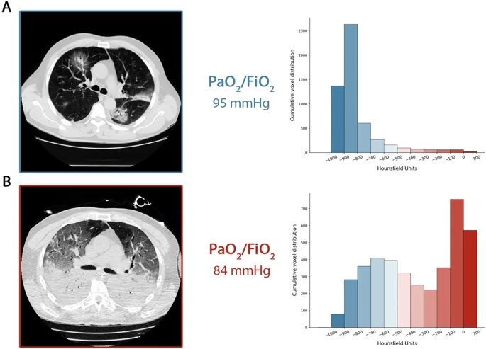 A CT scan acquired during spontaneous breathing. The cumulative distribution of the CT number shifts to the left (well-aerated compartments), with the compartment from 0 to - 100 HU, the non-aerated tissue being practically 0. In fact, the total weight of the lung tissue was 1108 g, 7.8 % that was not aerated and the gas volume was 4228 ml. Patient receiving oxygen with inspired venturi mask oxygen fraction of 0.8. b CT acquired during mechanical ventilation at end-expiratory pressure at 5 cmH2O PEEP. The cumulative distribution of the CT scan shifts to the right (unaerated compartments), while the left compartments are significantly reduced. In fact, the total weight of lung tissue was 2744 g, of which 54% was unaerated and the gas volume was 1360 ml. The patient was ventilated in controlled volume mode, 7.8 ml/kg tidal volume, respiratory rate of 20 breaths per minute, fraction of inspired oxygen of 0.7
Respiratory treatment
Given this conceptual model, it follows that the respiratory treatment offered to type L and type H patients must be different.
The proposed treatment is consistent with what has been observed in COVID-19, although the overwhelming number of patients seen in this pandemic may limit its broad applicability.
1) The first step to reverse hypoxemia is through an increase in FiO2 to which the type L patient responds well, particularly if they do not yet have dyspnea.
2) In type L patients with dyspnea , there are several non-invasive options available:
- High Flow Nasal Cannula (HFNC)
- Continuous positive airway pressure (CPAP)
- Non-invasive ventilation (NIV).
At this stage, measurement (or estimation) of oscillations of inspiratory esophageal pressure is crucial [13]. In the absence of esophageal manometry, surrogate measures of the work of breathing, such as central venous pressure changes [14] or clinical detection of excessive inspiratory effort, should be evaluated.
In intubated patients , P0.1 and P occlusion should also be determined . High PEEP, in some patients, can decrease pleural pressure changes and stop the vicious cycle that exacerbates lung injury. However, high PEEP in normally compliant patients may have detrimental effects on hemodynamics. In any case, non-invasive options are questionable, as they may be associated with high rates of failure and late intubation , in an illness that generally lasts several weeks.
3) The magnitude of the oscillations of inspiratory pleural pressures can determine the transition from Type L to Type H phenotype . As esophageal pressure changes increase from 5 to 10 cmH2O, which are generally well tolerated, to greater than 15 cmH2O, the risk of lung injury increases and therefore intubation should be performed as soon as possible.
4) Once intubated and deeply sedated, Type L patients, if hypercapnic , can be ventilated with volumes greater than 6 ml/kg (up to 8–9 ml/kg), as high compliance results in tension tolerable without risk of ventilation-induced injury (VILI). Prone positioning should be used only as a rescue maneuver, since lung conditions are "too good" for the effectiveness of the prone position, which is based on stress improvement and tension redistribution. PEEP should be reduced to 8-10 cmH2O, as recruitment capacity is low and the risk of hemodynamic failure increases at higher levels.
Early intubation can prevent the transition to the type H phenotype.
5) Type H patients should be treated as severe ARDS, including higher PEEP, if compatible with hemodynamics, prone positioning and extracorporeal support.
|


